-
-
-
400-685-8537

source: 仁树医疗 2026-07-31 17:49:24 Secondary reading
A car accident half a year ago changed the life of 2-year-old Xuanxuan (pseudonym) and his family. Since then, clear, watery fluid has been intermittently dripping from his nostrils—flowing when he stands up and stopping when he lies down.
His parents initially thought it was just a cold, but the "runny nose" persisted for days—dripping nonstop, with no sign of improvement. When they took him to the hospital, the diagnosis left the whole family stunned—cerebrospinal fluid (CSF) rhinorrhea. In plain terms: his brain was "leaking."
What made matters even more devastating was that after visiting multiple top-tier tertiary hospitals, they received only one answer—craniotomy.
At just 2 years and 10 months old, the child's brain was still in a phase of rapid development. A craniotomy meant incising the scalp, sawing through the skull, and retracting brain tissue. Both of Xuanxuan's parents were healthcare professionals themselves—they understood better than most what a craniotomy truly entailed: trauma, risks, a prolonged recovery, and the unknown consequences it might have on a developing brain.
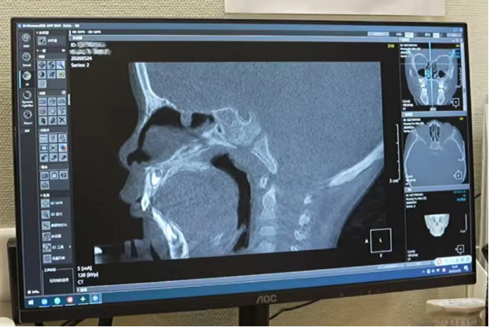
It wasn't until they came to Renshu Eye, Ear, Nose & Throat Hospital and met Professor Zuo Kejun that a different path emerged. After reviewing the CBCT 3D reconstruction images, Professor Zuo said: "The lesion can be reached through the nasal cavity—no craniotomy is needed."
Examinations, results, and treatment plan were all completed on the same day. The departments of anesthesiology, radiology, operating room, and nursing worked in rapid coordination—launching an efficient, multidisciplinary effort focused on diagnosing and managing an occult cerebrospinal fluid leak in a young child.
01 Why did multiple hospitals recommend craniotomy, while Renshu could offer a minimally invasive alternative?
What made Xuanxuan's case so unique? Two factors: his young age and small body size, combined with a tiny, elusive leak site.
At just 2 years and 10 months old, the operative space within the nasal cavity was measured in millimeters. The adult nasal cavity is already narrow enough; a child's is even smaller—small nostrils, a constricted nasal passage, leaving virtually no room for instrument manipulation once the endoscope is inserted. Moreover, the skull base defect measured only 1–2 mm in diameter. Precise localization under such conditions posed a considerable technical challenge—and was also the prerequisite for a successful surgery.
The traditional craniotomy repair approach follows a "top-down" strategy—finding the leak site through the skull—which requires incising the scalp, sawing through the skull, and retracting brain tissue. It is associated with significant trauma, high risks, a slow recovery, and a hospital stay of up to 21 days.
In contrast, Renshu transformed this craniotomy into a minimally invasive endoscopic endonasal procedure through six innovative techniques, enabling the child to be discharged in just 3 days.
02 Six Innovative Technologies: Breakthroughs from Diagnosis to Recovery
High-Resolution CT 3D Reconstruction: Accurate Localization in 15 Minutes
Leveraging high-resolution cone-beam CT (CBCT) and high-definition 3D reconstruction imaging, Professor Zuo precisely pinpointed the tiny 1–2 mm leak site. The examination and results were all completed on the same day—within just 15 minutes, with no lengthy waiting period. Compared with MRI, high-resolution CT provides clearer and more intuitive visualization of bony defects, offering "precision guidance" for the surgery.
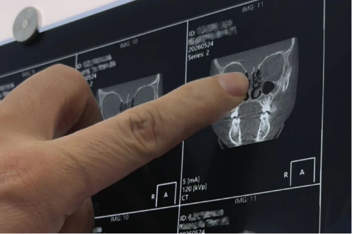
Efficient Collaboration: The radiology department synced the reconstruction data to the surgical team in real time, enabling seamless coordination from examination to diagnosis.
"Cold Instruments" for Creating the Graft Bed: Preserving Tissue Viability Without Electrocautery
During the procedure, fine instruments—referred to in the field as "cold instruments"—were used to meticulously prepare the area around the fistula. Without resorting to electrocautery or other thermal tools, the viability of normal tissues surrounding the fistula was maximally preserved, creating the optimal "soil" for subsequent healing and growth. This is the critical first step in ensuring rapid post-repair recovery.
Embedding Autologous Bone Chips via the Intracranial Approach: Turning Large Holes into Small Holes, and Small Holes into Cracks
Autologous bone fragments generated from the patient's own sinus during the surgery were harvested and trimmed into patches of appropriate size and shape. Using an intracranial embedding technique, the bone chips were inserted between the skull base and the dura mater, securely wedging into the leak site.
This approach effectively turns large defects into smaller ones, and small defects into mere cracks, reducing the outward flushing force of cerebrospinal fluid that might otherwise displace the extracranial repair layer. This maneuver demands extreme precision—if the bone chip is too large, it cannot be inserted; if too small, it will not hold securely. It must fit within a hairbreadth's margin, as this directly determines the long-term reliability of the repair.
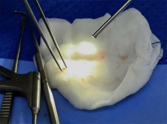
Pedicled Mucosal Flap: Designed Specifically for "Uncooperative" Young Children
Young children cannot be expected to comply with strict postoperative bed rest—crying and rolling around may easily displace the graft. Traditional methods fall short in addressing this issue. Professor Zuo pioneered the pedicled turbinate mucosal flap technique:
Precisely resecting the lower half of the superior turbinate, then elevating and extending the superior turbinate mucosa to form a living tissue flap with an intact blood supply, which is laid over the bone chip (extracranially);
For the dural defect in between, a free mucosal flap is fashioned into a fascial graft with rough surfaces on both sides to serve as the dural repair material;
The pedicled flap provides a continuous blood supply, ensuring graft stability and rapid healing.
With adequate vascularization, the repair time is significantly shortened, antibiotic use is reduced accordingly, and the risk of infection is minimized.
Absorbable Packing Material: No Need for Nasal Pack Removal Postoperatively
Traditional procedures require non-absorbable packing, which must be painfully removed after a few days—an experience that is difficult even for adults, let alone a 2-year-old child.
At Renshu, absorbable packing material was used for this young patient, eliminating the need for any secondary removal procedure, as the material is naturally absorbed and degraded by the body. After surgery, Xuanxuan woke up calm and quiet—largely attributed to this innovation.
Shortened Hospital Stay: From 21 Days to 3 Days
Traditional craniotomy repair requires a 21-day hospital stay. Even with the widespread adoption of endoscopic endonasal techniques, most hospitals still require 7–8 days.
Through Professor Zuo's triple-protection technique, combined with Renshu's meticulous perioperative management, the hospital stay was compressed to just 3 days post-surgery. Xuanxuan showed improved mental status on the first day after surgery, engaged in mild activity on the second day, and was discharged home on the third day.
03 Behind the Efficient Surgery: Multidisciplinary Collaboration Is Key
Preoperative Phase
The radiology department delivered the report within 15 minutes. The anesthesiology and nursing departments completed their evaluations on the same day. The surgery was scheduled as the first case early the next morning to avoid prolonged fasting for the child.
Intraoperative Phase
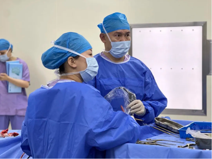
Renshu has a long-standing, dedicated anesthesia team and nursing partners who have developed a high level of默契 over years of collaboration. The anesthesiologist precisely calculated drug dosages tailored to the child's physiological characteristics—limited blood volume and poor thermoregulatory capacity—while closely monitoring vital signs throughout the procedure. Professor Zuo's continuous refinement of minimally invasive surgical techniques effectively shortened the operative time, thereby reducing anesthesia-related risks and minimizing surgical trauma.
Postoperative Phase
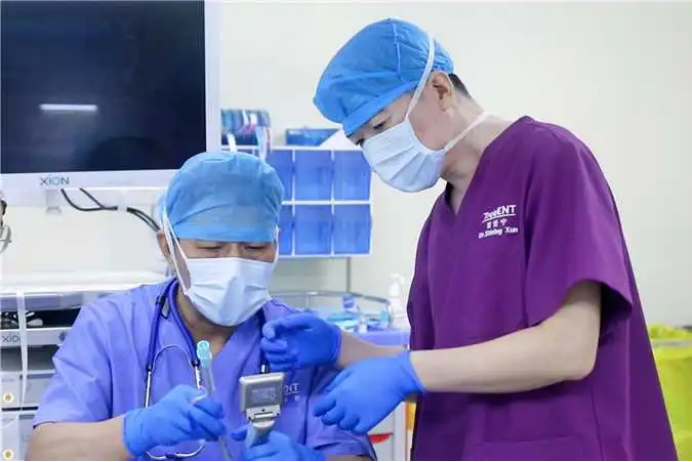
The nursing team provided specialized care—including postoperative observation, dietary guidance, medication management, and activity recommendations. After discharge, a 24-hour response mechanism was activated, allowing parents to reach out at any time with questions or concerns, ensuring a worry-free recovery process.
04 The Child Climbing onto the Examination Chair by Himself—The Best Answer to Medical Progress
Both of Xuanxuan's parents are healthcare professionals. They had seen too many postoperative patients—drowsy, in pain, crying and restless. But when Xuanxuan woke up after surgery, he was calm and quiet, with no crying or fussing. His mental state was even better than before the procedure, and his recovery far exceeded expectations.
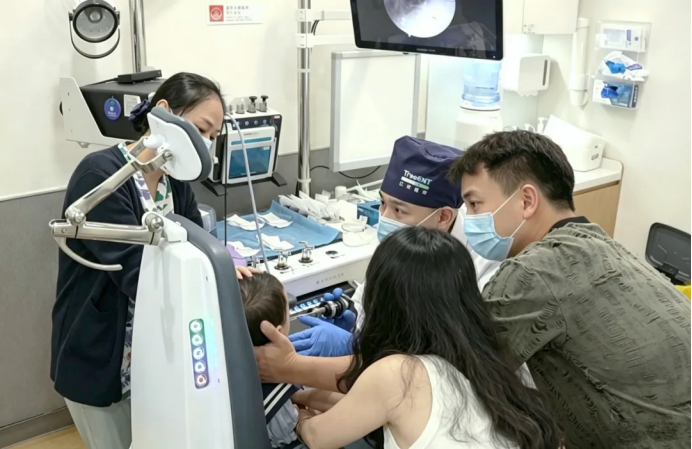
At the follow-up visit more than a month after surgery, the little boy climbed onto the examination chair all by himself, without a trace of fear. Professor Zuo smiled and said, "He's recovering beautifully—no need to come back anymore." His parents' eyes welled up with tears at the side. Half a year ago, they were still agonizing over whether to proceed with a craniotomy; half a year later, their child was already running around the clinic.
This, perhaps, is the most tangible meaning of medical progress for a family.
Important Reminder: Do NOT Delay!
If a diagnosis of cerebrospinal fluid rhinorrhea has been confirmed, timely intervention is absolutely critical—do not delay.
If left untreated for a prolonged period, the brain gradually adapts to the low-pressure "leaking" state. If surgery is postponed and the leak is eventually repaired, the sudden restoration of intracranial pressure may overwhelm the body's ability to adapt, potentially leading to serious complications such as intracranial hypertension.